By: Dr. Dev Batra | 01.29.23
Chronic vein disease develops in stages, and venous ulcers usually appear during the last and most serious stage. Because of poor circulation, open, shallow wounds form on the lower leg or ankle.
They’re slow to heal and can quickly become infected if not treated. That sometimes leads to lower-limb amputation, especially in diabetics. About 1% of Americans develop these ulcers, which are common in older people, especially women.
At Texas Vascular Institute, now with locations in Dallas and Hurst, Texas, interventional radiologist Dr. Dev Batra and his team have extensive knowledge about the stages of chronic vein disease and the development of venous ulcers.
The team uses state-of-the-art diagnostic tools to pinpoint the underlying cause of your issue and advanced treatment options to restore your vein health. Let’s take a closer look at venous ulcers.

How chronic vein disease progresses
Before we can talk about venous ulcers, we need to discuss the vein problems that cause them. Here are the stages of vein disease in order of progression.
1. Chronic venous insufficiency (CVI)
If a vein wall or one of the vein’s one-way valves becomes damaged, it allows blood headed for the heart to backtrack, pooling around the damaged area and contributing to sluggish flow. That venous insufficiency can easily become chronic if not addressed.
2. Spider veins
These surface-level veins are small, rope-like conduits. When blood pools, they expand, forming thin, spiderweb-like patterns on the skin, primarily on the legs but also on the face.
3. Varicose veins
As sluggish blood flow continues, the larger veins become affected. These varicose veins appear as ropy, colored swellings on the legs. In addition to being a cosmetic problem, they may cause pain, swelling, itchiness, and an aching heaviness in the legs.
4. Edema
Edema refers to the swelling of the leg when overloaded veins leak fluid into the tissues. In addition to causing swelling, the fluid irritates your muscles, which leads to cramping and pain as well as the symptoms of the previous stages. In addition, the skin on your lower legs may start to appear leathery or take on a pale blue or red tint.
5. Venous stasis dermatitis
If you don’t treat the edema, you may develop venous stasis dermatitis — a change in color around your ankles and lower legs that results from the pressure-bursting blood vessels. The areas appear brown or red due to hemosiderin deposits — a breakdown product of hemoglobin. The skin may also turn scaly, shiny, or thickened, and it may scar.


6. Venous ulcers
Venous ulcers occur when the skin changes from the dermatitis aren’t properly treated. Ulcers are easy to identify because they’re:
- Found on the lower leg or ankle area
- Surrounded by discolored and/or hardened skin with irregular borders
- Red at the base
- Slow to heal
Ulcers can produce large amounts of pus, depending on if they’re infected, and if you touch them, they ooze venous blood. The wound may be relatively painless, but you may experience significant pain from the edema or infection.
How to diagnose venous ulcers
Venous ulcers can easily be diagnosed based on sight. However, if Dr. Batra wants to confirm the diagnosis or discover the underlying cause, he might use vascular ultrasound and ankle-brachial index testing. Most commonly, he can trace the problem to chronic vein disease.
How we treat venous ulcers
At Texas Vascular Institute, our primary goal for treating venous ulcers is to keep them infection-free while also resolving the edema at the site. We may use debridement — scraping of dead tissue and surface contamination to remove it — so the ulcer can heal.
Generally, you’ll only need oral antibiotics if tissue surrounding the ulcer is also infected. We ask you to keep the wound environment moist, changing dressings as infrequently as possible since that removes healthy cells, too.
Most ulcers heal within 3-4 months of treatment, but it varies from person to person.
Dr. Batra also treats ulcers by addressing the underlying vein disease. Techniques include:
- VenaSeal™: delivers a medical glue to seal the diseased vein shut
- ClosureFast™: uses radiofrequency (RF) ablation to destroy the diseased vein
- Ultrasound-guided foam sclerotherapy: injects an irritant into the vein with ultrasound guidance
- Microphlebectomy: pulls out the entire vein through small incisions
If you’re experiencing any of the symptoms of vein disease, don’t wait until it’s advanced to get help. Call Texas Vascular Institute at 972-646-8346 to schedule a consultation with Dr. Batra at either location or book online with us today.
Read more blogs
Varicose Veins in Hurst: Expert Care at Your Doorstep
At Texas Vascular Institute (TVI), we empathize with the discomfort and worry caused by varicose veins. That's why we're here in Hurst, providing cutting-edge treatments that are customized to address your unique needs. With our team of experts wielding extensive knowledge and experience, we promise to provide the utmost care in a warm and compassionate atmosphere. Let us help you find relief and regain your confidence!
Varicose Veins in Dallas: Quality Care You Can Trust
Our exceptional team of vascular specialists are true leaders in their field, armed with years of invaluable experience. Harnessing the power of cutting-edge advancements in vein treatment, they've transformed the lives of numerous patients, liberating them from the pain and unsightly burden of varicose veins. When you choose TVI, you're opting for unparalleled care and unwavering commitment to your varicose vein needs in Dallas.
How to Get Rid of Varicose Veins in Hurst?
The causes and risk factors of varicose veins vary from genetics to age, pregnancy, obesity, and prolonged standing or sitting, among other factors. Some typical signs and possible issues include discomfort, inflammation, irritation, hemorrhage, dermatological alterations, sores, and thrombosis. You may want to seek medical attention if you experience any of the following symptoms or complications.
WHAT OUR PATIENTS
have to say
Texas Vascular Institute always appreciates feedback from our valued patients. To date, we’re thrilled to have collected 378 reviews with an average rating of 5 out of 5 stars. Please read what others are saying about Texas Vascular Institute below, and as always, we would love to collect your feedback.
Leave a Review
Amazing Practice
I'm very particular with my Healthcare and tend to be cautious with referrals to specialists. This office is amazing from the first point of contact. Their staff are friendly, professional and highly knowledgeable. Then the Dr is just as amazing as his staff, absolutely brilliant. Office manager Jessica has this office running like a well oiled machine and does so with a smile, an air of confidence, kindness and professionalism. Love this practice!!
- Richard G.

Beyond Thankful
Dr Batra and his staff are amazing! We are so grateful to have found him. Everyone is so kind and so caring and Dr Batra explains everything so well and does procedures with excellence. Beyond thankful to be under their care!!!
- Bitsy P.

Gold Standard
This is a gold standard for how a medical practice should be run. I was promptly seen at my scheduled time, my ultrasound was thorough and I received plenty of attention and care from the staff and Dr.Batra.
- Weronika L.
INSURANCE
We accept most major insurance plans. Please contact the medical office for all insurance related questions.


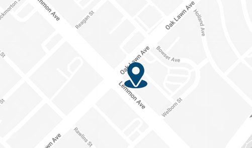
3500 Oak Lawn Ave, #760
Dallas, TX 75219
For Appointments: 972-798-4710
General Inquiries: 972-646-8346

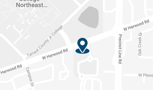
809 West Harwood Rd, Suite 101,
Hurst, TX 76054
For Appointments: 972-798-4710
General Inquiries: 972-646-8346

